
Two things drive me crazy, and a third vexes my soul:
- Over-diagnosis of subchorionic hematoma;
- Inappropriate counseling and recommendations regarding subchorionic hematoma;
- And inappropriate management of placental lakes.
Not a week goes by that I don’t see a patient who has presented to an emergency department somewhere, diagnosed with a (questionable) subchorionic hematoma or placental lake, and then essentially told to expect the worst and put on bed rest. It’s frankly amazing to me and frustrating because I am left to clean up the emotional trauma. So let’s review these diagnoses and their management.
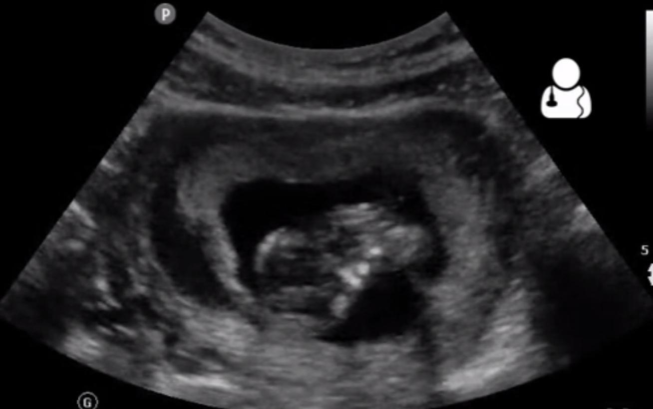
Subchorionic Hematoma or Hemorrhage.
A subchorionic hematoma typically appears as a sonolucent, crescent-shaped finding that potentially represents bleeding from a partial detachment of the trophoblast from the uterine wall. Blood then dissects between the chorion and the decidual layer of the endometrium.
The appearance of a subchorionic hematoma is variable. If there is active bleeding, then the collection of fluid may not be echogenic; as clot forms, it may become hyperechogenic; as clot resorbs and there is liquefaction, the echogenicity may decrease. The shape, too, can vary, from the typical crescent shape to ovoid or linear.
Over-diagnosis is very common. The reported incidence of a subchorionic hematoma ranges from 4-22%, a sign that there is significant disagreement about what constitutes a true subchorionic hematoma. In early pregnancy in particular, when many ER visits for bleeding or cramping occur, subchorionic hematomas can be incorrectly diagnosed for an incomplete chorioamniotic fusion or a vanishing twin. Both of these findings are not associated with adverse pregnancy outcomes.
Even when a true subchorionic hematoma is present, it should be graded according to size.
- Small hematoma (less than one third of the circumference of the GS – the hematoma in the picture is small)
- Medium hematoma (one third to one half the size of the GS)
- Large hematoma (at least two thirds the size of the GS)
This distinction is vitally important. The pregnancy loss rate ranges from 7.7% for small hematoma to over 18.8% for large hematomas. For moderate sized hematomas, the loss rate is about 9.2%. In other words, women with small and moderate hematomas are not at an increased risk of pregnancy loss compared to the average pregnant woman, and small and moderate hematomas (or misdiagnosis) accounts for most ultrasonographic diagnoses of hematomas.
The actual measured impact on miscarriage rate (and potential preterm delivery rate or abruption rate) varies widely in studies. Some data sets indicate no increased risk of miscarriage whatsoever, while others record significantly higher rates of miscarriage. The likely explanation of this difference is the size of the hematoma. Older data indicated higher rates of miscarriage, and this older data showed lower rates of subchorionic hematoma. In other words, the older data sets are more likely to contain symptomatic women (those who present with pain and bleeding) and larger hematomas (those bleeds more easily seen on 1990s-era ultrasound machines).
Newer data tends to shows higher rates of subchorionic hemorrhage, more often diagnosed in asymptomatic women (such as on routine dating scans). In this population of women, with more small and moderate size bleeds, the rate of miscarriage is not significantly different compared to women without hematomas. It is this latter group of patients that causes me the most anxiety, because many obstetricians (but particularly non-obstetricians) counsel the patients in the worst possible way.
Recall that even in the presence of a symptomatic patient with a large hematoma, the rate of miscarriage is still only about 1 in 5. Put another way, the patient sitting in front of you with bleeding and a large hematoma has an 81.2% chance that she will not miscarry. That’s roughly equivalent to the risk of miscarriage that a woman is exposed to when she has a positive test at home (before seeing cardiac activity). In other words, STOP SCARING WOMEN TO DEATH!
Too many women have stories like this momma, whose impression leaving her midwife’s office was that she should expect to miscarry. She was also told to be on bedrest, to reduce her stress level, to have someone else take care of her children, and to not eat (on the first day) in case she needed a D&C! Later, she learned that most subchorionic hemorrhages do okay online, but she also learned of the healing properties of watermelon for subchorionic hematoma in a support group; in others words, don’t leave your patients to learn information online – educate them properly.
I like reading stories from mothers who have blogged about their experiences. Most of the stories are simply embarrassing to Obstetrics. Here is another momma who reveals her fear and misguided counseling. In addition to being placed on strict bedrest, she was also under the impression that the chances of her not miscarrying and/or making it passed 20 weeks were “slim” at best. As is usually the case, she was induced at term and the hematoma was of no consequence.
Another theme in these stories is that most women are scared first by their primary OB/GYN, then they are referred to Maternal-Fetal Medicine. The finding of a subchorionic hemorrhage, even if large, doesn’t require referral. When a patient is referred, the MFM feels a therapeutic imperative, and this is the usual origin of advice like bedrest; he feels he must do something, and the MFM seldom wants to reassure the patient that there isn’t much to worry about or much to do because that tends to make the referring physician look incompetent. Worse, MFMs are all too happy to “follow” just about anything with ultrasound – it’s a cash cow, after all. But we should only “follow” things with serial testing if it might potentially change the management of the patient. This always reminds me of the common practice of repeating an ultrasound to see if choroid plexus cysts have resolved – it doesn’t matter!
How should a true subchorionic hemorrhage be managed?
As with many pregnancy conditions, there’s really nothing to do except to avoid making things worse. So,
- Don’t recommend activity restriction, including pelvic rest, stopping exercise, or bed rest. None of these strategies are associated with improved outcomes. All three can lead to guilt sanctioning for the mother who does go on to miscarry or have a bad outcome. It also financially harms patients and families. Bed rest will actually worsen physical outcomes, as it increases the pregnant woman’s risk for a variety of poor outcomes including death (due to thromboembolism).
- Don’t scare her with unsupported claims. In other words, if it is a small incidental finding, don’t even mention it. If it is a large hematoma in a symptomatic patient, give her real numbers: “This finding is associated with a slightly higher risk of miscarriage, but overall more than 80% of women like you go on to have a baby.”
- Don’t do unnecessary follow-up scans. There is no therapeutic and little diagnostic value in repeated ultrasounds to follow the hematoma. Don’t exploit the patient.
Placental Lakes
Another common ultrasound finding during pregnancy a lake or lacuna. This is common both during the first trimester and later in pregnancy. The incidence of these sonolucenies varies widely in studies, from about 2% to over 17% in the 2nd and 3rd trimesters. These lakes appear in the substance of the placenta, as opposed to the subchorionic hematomas that are between the chorion and uterine wall.
The presence of these lacuna are not associated with any adverse pregnancy outcomes when detected in the first trimester. First trimester diagnosis has become more common with the widespread performance of first trimester nuchal translucency ultrasounds. Too often, patients are cautiously told about these lakes and they are used as a justification for repeated ultrasounds. This is not indicated.
In the second and third trimesters, placental lakes may be associated with two things. Most important of these, the lakes, along with other findings, may be associated with invasive placental disease. However, suspicion of placenta accreta, increta, or percreta should be limited to those patients with appropriate history, e.g., previous Cesarean with a placenta overlying the scar.
Apart from this, large lakes (>5 cm) noticed in the second or third trimester have been associated with IUGR. It seems reasonable to follow pregnancies with large lakes with ultrasound. But, as is the case with subchorionic hemorrhage, only the large ones seems to matter and the large ones are very rare.
In general, the same management advice applies: don’t scare women to death, don’t do unnecessary follow-up ultrasounds (unless its very large), and don’t recommend other nonsense like bedrest.