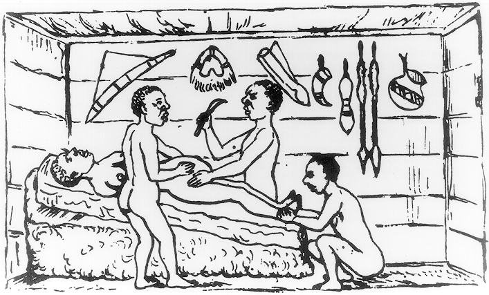
There are billions of ways to do a cesarean delivery. At least 203,843,174,400 ways that I know of (well that’s how many we are going to talk about in this post anyway).
One of my mentors used to say, “Surgery is a thousand little things done well,” and it’s very true. We make hundreds of little decisions every time we do a surgery, with minor, and sometimes major, differences in technique. Many of these differences are not evidence-based (that is, there simply is no evidence available). Many of the differences in technique are meaningless. Many are very important with good evidence behind them.
When surgery goes well, it often does so in spite of many things that could have been done better. When it goes poorly, many good techniques are often blamed. Bias pervades surgical technique, and many techniques or habits persist like good luck charms in a baseball player’s pocket. Dogma is entrenched in the operating room (we will discuss this more at the end of the Post).
Let’s go through and look at some of the major decisions/techniques that face the obstetrician during a cesarean delivery. I will place in bold the choices I favor and where good evidence is available, I’ll provide a link. Admittedly, there isn’t data available for everything.
Where available, I have included the USPTFS strength of recommendation and level of evidence (please review these, but basically A and B indicate that you should do it, C indicates that it may be appropriate in some cases, D indicates you should not do it, and I indicates insufficient data).
Most, but not all, of the Evidence links below point to Evidence-based surgery for cesarean delivery: an updated systematic review, which is well-worth reading.
Universal administration of antibiotics
- Before case starts (Evidence, A-High)
- After baby is born
Hair prep
- Clip (Evidence)
- Shave
Skin prep
- Chlorhexadine (Evidence, I-Low)
- Betadine
Drape
- Ioban drape (Not studied in obstetrics, but some compelling data in other specialties)
- Normal drape
Vaginal prep
- Preoperative douche of the vagina (Evidence, B-Moderate)
- No preop douche
Tilt
- Left lateral tilt of the patient
- No tilt (Evidence, I-Low)
Skin incision
- Pfannenstial
- Joel-Cohen (Evidence)
- Maylard
- Vertical
- Paraumbilical
Cut down method through fat and knicking the fascia
- With Bovie
- With scalpel (Evidence for and Evidence against) (This is almost a philosophical question, which we will talk about at the end)
Fascial incision extension
- Sharp dissection of the fascia
- Blunt dissection of the fascia (not studied individually, but part of overall techniques, such as Misgav-Ladach cesarean or the Joel-Cohen incision, which have been found to be superior in numerous trials. Again, see below)
Dissection of the rectus muscles
- Dissect rectus muscles off the fascia (with scissors or Bovie)
- Do not dissect muscles off the fascia (Evidence, I-Low, RCT)
Peritoneal incision
- Sharp creation and extension of incision
- Blunt creation and extension (see below)
Bladder flap
- Create a bladder flap (Evidence, D-Moderate, Meta-analysis)
- Don’t create a bladder flap
Scalpel
- Same scalpel as used on skin
- Different scalpel as used on skin
Uterine incision
- Low transverse (Evidence)
- Low vertical
- Classical
Uterine extension
- Sharp (with scissors after scalpel)
- Blunt in a bilateral direction
- Blunt in a cephalocaudad direction (Evidence, Meta-analysis, My commentary)
Delivery of the baby (if difficulty is encountered)
- Difficult delivery with forceps (no evidence)
- Difficult delivery with vacuum extractor
Delivery of the placenta
- Massage of the uterus (Credé) with traction (Evidence, A-High, Cochrane Review)
- Await spontaneous expulsion (also certainly acceptable)
- Manual extraction
Manual exploration of the uterus
- Clean out the uterus with a sponge
- Don’t clean out the uterus with a sponge (Evidence, I-Low)
Cervix dilation
- Mechanical dilation of the cervix to allow drainage (Evidence, D-Moderate)
- Non-dilation
Uterine exteriorization
- Exteriorization during repair (Evidence, C-Moderate)
- Nonexteriorization
Myotomy repair style (there are obviously many other choices than those listed below)
- Single layer, running, locked (Evidence, A-High for women not planning pregnancy, )
- Single layer, running, not locked
- Double layer with second layer imbricating
- Double layer with second layer nonimbricating
- Interrupted figures of 8
Incorporation of peritoneal edges in myotomy repair
- Incorporation (Evidence, I-Low)
- Nonincorporation
Myotomy suture material
- Vicryl
- Chromic (no good data, some animal studies)
- Monocryl
Vesicouterine peritoneal closure
- Closure
- Nonclosure (Evidence, C-Moderate)
Irrigation
Abdominal peritoneal closure
- Closure
- Nonclosure (Evidence, C-Moderate, Cochrane Review)
Rectus muscles
- Reapproximation
- Nonapproximation (generally data is included in peritoneal closure studies)
Fascial closure
- Vicryl
- One suture (for primary sections)
- Two (one from each angle meeting in the middle) (doubles risk of knot failure)
- PDS (for repeat sections or morbidly obese)
Subcutaneous closure
- Approximation of tissue when deeper than 2 cm (Evidence, A-High)
- Nonapproximation
Placement of drain
- Placement of drain when tissue is deeper than 2 cm (Evidence, D-High)
- No placement of drain
Skin closure
- Subcuticular suture (Evidence)
- Skin staples
Dressing
- Glue (Evidence)
- Bandage
So that gets us to 203 billion combinations, and obviously there are many more options than we have discussed. We could get to a trillion just by talking about three more things. That large number should demonstrate one of the inherent problems in studies that involve surgical technique: it takes a large number of procedures to show minor differences in outcomes and there are many, many variables to be accounted for. We haven’t even considered the variability in patients.
Nevertheless, some techniques can be studied sufficiently to determine that they should be done, and those are things represented above by ‘A’ and ‘D’ evidence: universal antibiotic prophylaxis before the case starts, blunt cephalocaudad extension of the uterine incision, non-manual removal of the placenta, one layer closure of the uterus if no future fertility is desired, subcutaneous closure if ≥2 cm fat, no irrigation, no placement of drains, and no manual dilation of the cervix.
Other things are based on lower level data; that doesn’t mean that the options are equal, it just means we are less sure. This includes things like non-closure of the peritoneum and subcuticular closure of the skin. And of course several items need more study. Some, like double layer uterine closure for women who desire future fertility, will likely never be answered in a clinical trial. But that difficulty in designing a trial to answer the question also shows us that it probably just doesn’t matter one way or the other, like exteriorization of the uterus.
Here is a presentation that reviews much of this data with more explanations from obgynstudent.com.
Now the philosophical portion. The classical technique for Cesarean Delivery, and most surgery, has its roots in the surgical principles of Halstead, which include:
- Gentle handling of tissue
- Meticulous hemostasis
- Preservation of blood supply
- Strict aseptic technique
- Minimum tension on tissues
- Accurate tissue apposition
- Obliteration of deadspace
These dogmas have become beaten into the minds of surgeons, as if they were delivered from Mount Sinai. Unfortunately, they are not all scientifically accurate. And several extensions of these principles have also become dogma, like, “Dilution is the solution to pollution,” which advocates for routine irrigation.
In a Halsteadian cesarean delivery, we would cut each layer of tissue sharply and meticulously rather than pulling and tearing. We would close each of these layers back carefully, including all layers of peritoneum and the muscles. We would irrigate both the peritoneal cavity and the abdominal incision. We would cauterize all the bleeding vessels we could find, including the inferior epigastrics (even though this is at odds with Principle 3).
But Halstead is at odds with 100 years of surgical progress. Minimally invasive surgery emphasizes small incision and minimal disruption of blood vessels and tissue. It emphasizes placing as few foreign bodies (sutures) in the patient as possible since these are a nidus for inflammation. Its tries to preserve blood supply, even small vessels, by not cauterizing and devitalizing tissue unnecessarily.
In a minimally invasive approach, we enter tissues bluntly and stretch them, preserving more intact anatomy and minimizing blood loss and post-operative pain. We use less sutures, not out of laziness, but out of a recognition of how the body actually heals (the peritoneum for example) and out of desire to reduce inflammation and associated pain and adhesions.
Even though a Cesarean Delivery can hardly feel like a minimally invasive procedure, still, its invasiveness can be minimized. This was the basis of the Joel-Cohen skin incision, and the Misgav-Ladach cesarean (also called Stark Procedure). These “modern” techniques (from the 1960-70s) have been built-upon and improved, but they represent a departure point from older, Halsteadian and antiquated techniques.
Unfortunately, most obstetricians have not incorporated even the highest-level evidence-based techniques into their practices. This has more to do with cognitive bias than anything else, which will be a frequent topic of this blog.

