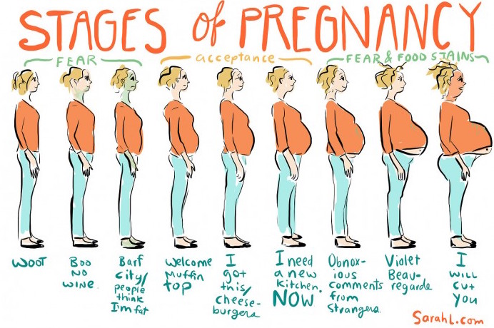
From SarahL.com
Much of what constitutes prenatal care is based on tradition and lore. Having a clear understanding of evidence-based practices is important. For the healthy, low-risk pregnant woman, most routine interventions are unnecessary and potentially harmful. For at-risk patients, prenatal care should be individualized. In many cases, the most effective part of prenatal care is education and emotional support.
How often should visits be?
Ideally, the first prenatal visit occurs in the first trimester. Of course, many women may present for care much later. Traditionally, visits after the first visit occur monthly until 28 weeks, then twice-monthly until 36 weeks, then weekly. This visit frequency is certainly not rigid. Many women may require more frequent visits depending upon on-going problems and/or risk-factors; conversely, many women may require fewer visits. Thus, the visit frequency should be individualized for each patient, also taking into consideration her anxiety level, etc.
Our traditional model of prenatal visits was built around screening for hypertension almost 100 years ago, with more frequent visits in the third trimester as the incidence of hypertension increases. But for many patients at low risk, especially in the age of ambulatory blood pressure monitoring, less frequent visits at the end may be desirable. Patient satisfaction tends to be higher with fewer visits, and less frequent visits at the end may decrease the number of unnecessary inductions.
What labs or tests are important?
- Prenatal labs.
- Rubella immunity status.
- Screening for anemia with a CBC (including evaluation of the MCV).
- STIs/STDs:
- Chlamydia
- Gonorrhea
- Hepatitis B
- HIV
- Syphilis
- Blood type and antibody screening;.
- Diabetes screening if patient at high risk
- In higher risk populations, HCV and urine drug screening are often appropriate.
- Screen by history for maternal thyroid disease. High risk women or women with a history of thyroid disease should receive a TSH with reflex free T4.
- Ultrasound.
- Ultrasound may be performed at the first prenatal visit to confirm gestational age, viability, fetal number, and fetal location. Fetal cardiac activity can be seen as early as 5 weeks and 5 days by transvaginal ultrasound.
- A routine anatomic survey should be performed at 18-20 weeks.
- Follow-up ultrasounds should be performed as indicated.
- Serial growth ultrasounds on patients at risk for fetal growth abnormalities:
- Diabetes, renal disease, autoimmune disease, cyanotic heart disease, hypertensive disorders, antiphospholipid antibody syndrome, substance abuse, multiple gestations, certain teratogen exposures, certain infectious disease exposures.
- Antepartum testing (possibly with biophysical profiles or nonstress testing) for at risk patients:
- Maternal conditions: pregestational diabetes, hypertension, SLE, renal disease, antiphospholipid antibody syndrome, poorly-controlled hyperthyroidism, hemoglobinopathies, cyanotic heart disease.
- Pregnancy-related conditions: gestational hypertension, preeclampsia, decreased fetal movement, poorly controlled or medically controlled gestational diabetes, oligohydramnios, fetal growth restriction, late term or post-term gestations, isoimmunization, previous fetal demise, monochorionic twins.
- Serial growth ultrasounds on patients at risk for fetal growth abnormalities:
- Fetal aneuploidy screening. There are many overlapping and confusing options available for screening for fetal aneuploidy. Women should be counseled about available options and make an informed decision. As with any genetic-type testing, patients should understand the consequences of testing and have a clear understanding of what they intend to do with positive results. Many well-informed women will choose no screening at all. Options include:
- First trimester screening (nuchal translucency, PAPP-A, and HCG) performed between 11-13 weeks gestation.
- Cell-free DNA testing or NIPT (non-invasive prenatal testing) performed any time after 10 weeks gestation.
- Quad or Tetra screen (AFP, hCG, estriol, inhibin A) performed 15-20 weeks gestation.
- Specific genetic testing. This should based upon the ethnic or genetic history of the patient and/or her partner.
What things should be done at each visit?
- First prenatal visit.
- Establish accurate dating.
- Detailed history, including screening for risk factors affecting pregnancy.
- Directed physical exam.
- Screen for intimate partner violence, depression, and other mental health or social issues which might affect pregnancy.
- Draw prenatal labs.
- Pap smears are often performed at the first prenatal visit but are seldom necessary. Pap screening should occur only if it happens to be due per normal screening recommendations.
- Counsel regarding treatments for common bothersome symptoms of pregnancy, including nausea, vomiting, constipation, etc. Provide patient with a list of “safe” medications to take for common ailments.
- 12 weeks gestation.
- Pregnant women should be screened for asymptomatic bacteriuria between 11-16 weeks gestation to decrease the risk of recurrent UTI, pyelonephritis, and preterm labor. There is no evidence to support screening at other times.
- First trimester screening if desired.
- 18-20 weeks gestation.
- Anatomic ultrasound.
- Quad screen if desired.
- 24-28 weeks gestation.
- Screening for gestational diabetes by history and, in many cases, by 1-hour, 50 g, glucose challenge test (GCT).
- Administration of RhoGam at 28 weeks.
- In high risk patients, may repeat anemia screening.
- 32 weeks gestation.
- TDaP vaccination (between 27-36 weeks gestation)
- 36 weeks gestation.
- Confirm fetal presentation.
- Encourage external cephalic version if non-cephalic.
- Group B Strep (GBS) screening (performed within 5 weeks of latest anticipated date of delivery) unless patient has prior history of GBS affected infant or a GBS positive urine culture in current pregnancy.
- In high risk patients, may repeat HIV screen.
- Plan for repeat caesarean delivery, if desired, at 39 weeks gestation.
- Confirm fetal presentation.
- 40+ weeks gestation.
- Schedule delivery no later than 41 weeks gestation.
In general, the following should happen at each prenatal visit:
- Maternal weight measurement.
- Blood pressure measurement.
- Abdominal palpation (at 36 weeks or greater) to determine fetal position and estimate fetal size.
- Doppler of the fetal heart tones (typically at each visit after 10-12 weeks). This does not change fetal outcomes but is reassuring to the mother and confirms continuing viability.
- Urinalysis for asymptomatic bacteriuria between 11-16 weeks. Urine dipstick at any visit in which the patient is hypertensive. Urinalysis at other visits lacks evidence.
- Measurement of fundal height is traditionally performed, but due to intra- and inter-observer variability, data has failed to demonstrate any benefit. Screening for growth abnormalities should be based on risk factors and third trimester palpation.
- History should focus on signs of preterm labor (vaginal bleeding, contractions/cramping, leaking of fluid), fetal movement (after the mother first reports quickening, usually around 20 weeks), and specific complaints.
A graphic representation of prenatal visits is available here from obgynstudent.com.
What are some important interventions?
- Prenatal vitamins. Folic acid supplementation prenatally should be universally recommended to prevent neural tube defects. However, the neural tube closes at 28 days post-conception, so evidence of the utility of folate supplementation after this is lacking. As far as the other components typically found in prenatal vitamins, there is scant data of efficacy or necessity. There are some retrospective studies which indicate that prenatal vitamins may decrease the risk of preeclampsia in underweight and normal weight women, as well as decrease the risk of preterm labor. These effects were not observed in overweight or obese women. The reduction in risk of preeclampsia seems of value only if taken 3 months pre-conceptually. The bottom line: there is no good data which indicates that prenatal vitamins benefit women with normal diets, apart from folate supplementation pre-conceptually (review article). Therefore, it is important to emphasis to women planning or capable of pregnancy to supplement folic acid.
- Iron supplementation. Iron supplementation, whether in a prenatal vitamin or separately, is only needed for women who are known to be iron deficient. No known benefits exist for most women and overall iron supplementation may pose potential harm (at the very least, increased constipation).
- Aspirin. Women who are at high risk for preeclampsia benefit from 81mg ASA supplementation after 12 weeks of gestation (with some evidence that it is beneficial before then). Those at high risk for preeclampsia are women with any one of the following:
- History of preeclampsia, especially when accompanied by an adverse outcome
- Multifetal gestation
- Chronic hypertension
- Type 1 or 2 diabetes
- Renal disease
- Autoimmune disease (systemic lupus erythematous, antiphospholipid syndrome)
- Rh(D) Immune Globulin (RhoGam). All Rh-negative women should be given RhoGam at 28 weeks gestation and/or at anytime there is significant bleeding or risk of feto-maternal hemorrhage (such as trauma, amniocentesis, external cephalic version).
- Influenza vaccination. All pregnant women should receive the influenza vaccine during flu season at the first opportunity. Other immunization-specific information can be found at immunizationforwomen.org.
- 17-hydroxyprogesterone caproate (Makena). Women with a prior spontaneous preterm birth should be given weekly injections of Makena starting at 16 weeks until 36 weeks to reduce the risk of subsequent preterm birth.
What are important educational topics?
- Smoking cessation.
- Substance abuse.
- Breastfeeding education and encouragement.
- Availability of childbirth education classes.
- Exercise. Advise at least 30 minutes of moderate exercise most days per week. Most restrictions on exercise are anecdotal. There is no evidence, for example, that women should avoid a maximum heart rate. All types of exercise are acceptable but women should be cautioned to avoid high impact sports (due to blunt trauma risk) and that they are more prone to orthopedic injury and accidents. Pregnant women should also avoid SCUBA diving.
- Preterm labor signs and symptoms. No interventions exist to successfully abort preterm labor. However, early identification is important to provide a window of opportunity for administration of corticosteroids and, if necessary, transfer to a facility with an appopriate NICU. Therefore, patients should be counseled in detail about signs and symptoms of preterm labor.
- Safety of medications, supplements, and potential (often work-related) environmental exposures. Known teratogens are rarer than many patients and providers might estimate. A detailed history of medications and supplements, as well as work exposures, should be performed for each patient. A useful resource for checking exposures is mothertobaby.org.
- Counseling on risks and benefits of a TOLAC. Rates of trials of labor after cesarean delivery (TOLAC) remain unjustifiably low. Patients should receive detailed counseling about the risks and benefits of a TOLAC versus a repeat cesarean delivery. Patients should be counseled about individual chances of a successful VBAC, which can be calculated here.
- Diet. Very few dietary recommendations have an evidence basis.
- Methylmercury. Pregnant women should avoid shark, swordfish, king mackerel, and tilefish.
- Listeria. Pregnant women are traditionally advised to avoid uncooked hot dogs and luncheon meats, or unpasteurized soft cheeses, to avoid listeriosis. However, recent listeria outbreaks have occurred with lettuce and other vegetables, including frozen vegetables, ice cream, hummus, and other products. The absolute risk of listeriosis from food stuffs is incredibly low and comparable to the risk of dying in a car accident while driving to prenatal visits. Providers should individualize this advice. In general, avoiding unpasteurized products in general as well as heating meats seems reasonable for most patients.
- Pregnant women should avoid supplementing excess iodine, as well as Vitamins A, D, K, and E.
- Few scientific studies are critical of caffeine in pregnancy. Based on relatively poor quality data, a recommendation of avoiding more than 350 mg of caffeine per day while pregnant has been made.
What things should we not do?
- Fetal kick count movements. Counting fetal movement should not be recommended to pregnant women. There are no improved outcomes with the practice, but significantly increased maternal anxiety, triage visits, and interventions.
- Bed rest. Both the American Congress of Obstetricians and Gynecologists and the Society for Maternal Fetal Medicine specifically recommend that bed rest should not be prescribed to pregnant women, even women at high risk for preterm labor, or other conditions. No studies have demonstrated benefit for bed rest or diminished activity for pregnancy complications, while multiple studies have demonstrated harm.
- Screening for bacterial vaginosis. Routine universal screening for BV does not decrease the risk of delivery before 37 weeks.
- Screening for HSV, Trichomonas, or Condylomata. Screening for HSV should be by history, and women with a history of HSV lesions should receive chemoprophylaxis in the last month of pregnancy.
- Screening for toxoplasmosis, cytomegalovirus, or parvovirus. A large portion of the population have serum antibodies to these pathogens, and no interventions exist that justify screening.
- Routine digital examination. No clinical value exists for routine digital exams of the cervix, and the uncomfortable practice may increase patient anxiety and complaints of bleeding, etc.
- Pelvimetry. There is no value in routine pelvimetry and provider comments about pelvis size may serve as discouragement for women about their ability to delivery vaginally.
- Routine evaluation for edema. Edema occurs in 80% of pregnant women and is too nonspecific to have clinical utility.
- Routine third trimester ultrasound for fetal weight. No studies have shown utility for this common practice, and these ultrasounds may encourage over-utilization of cesarean delivery for suspected fetal macrosomia.
For further reading:

